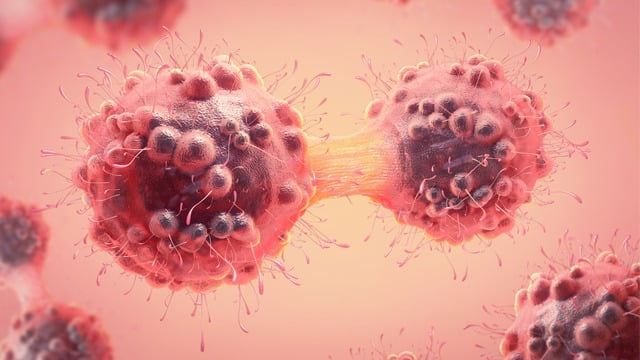
Washington: Researchers discovered an artificial intelligence system that could help pathologists read biopsies more accurately and better detect and diagnose breast cancer. The new system, described in a study published in the journal ‘JAMA Network Open,’ helped interpret medical images used to diagnose breast cancer that can be difficult for the human eye to classify. It does so nearly as accurately or better as experienced pathologists. It is critical to get a correct diagnosis from the beginning so that we can guide patients to the most effective treatments,” said Dr. Joann Elmore, the study’s senior author and a professor of medicine at the David Geffen School of Medicine at UCLA. A 2015 study led by Elmore found that pathologists often disagree on the interpretation of breast biopsies, which are performed on millions of women each year.

That earlier research revealed that diagnostic errors occurred in about one out of every six women who had ductal carcinoma in situ (a noninvasive type of breast cancer) and that incorrect diagnoses were given in about half of the biopsy cases of breast atypia (abnormal cells that are associated with a higher risk for breast cancer). Medical images of breast biopsies contain many complex data, and interpreting them can be very subjective. Distinguishing breast atypia from ductal carcinoma in situ is important clinically but very challenging for pathologists. Sometimes, doctors do not even agree with their previous diagnosis when they are shown the same case a year later,” said Elmore, who is also a researcher at the UCLA Jonsson Comprehensive Cancer Center.
The scientists reasoned that artificial intelligence could provide more accurate readings because by drawing from a large data set, the system can recognize patterns in the samples associated with cancer but are difficult for humans to see. The team fed 240 breast biopsy images into a computer, training it to recognize patterns associated with several types of breast lesions, ranging from benign (noncancerous) and atypia to ductal carcinoma in situ, or DCIS, and invasive breast cancer. Separately, the correct diagnoses for each image were determined by a consensus among three expert pathologists.
TTheresearchers compared its readings to independent diagnoses made by 87 practising U.S. pathologists. To test the system While the artificial intelligence program came close to performing and human doctors in differentiating cancer from non-cancer cases, the AI program outperformed doctors when differentiating DCIS from atypia, considered the greatest challenge in breast cancer diagnosis.
The system correctly determined whether scans showed DCIS or atypia more often than the doctors; it had sensitivity between 0.88 and 0.89, while the pathologists’ average sensitivity was 0.70. (A higher sensitivity score indicates a greater likelihood that diagnosis and classification are correct.) These results are very encouraging. There is low accuracy among practicing pathologists in the U.S. regarding the diagnosis of atypia and ductal carcinoma in situ, and the computer-based automated approach shows great promise.
 Fit Netion My WordPress Blog


